| Strain Name |
NOD.CB17-Prkdcscid Il2rgtm1Bcgen
Sirpatm1(SIRPA)Bcgen Cd47 tm1(CD47)Bcgen/Bcgen
|
Common Name |
B-NDG hSIRPA/hCD47 mice |
| Background | B-NDG | Catalog number | 110603 |
|
Related Genes |
SIRPα (signal regulatory protein alpha) CD47 |
||
|
NCBI Gene ID |
19261,16423 | ||
Mice Description
Signal regulatory protein α (SIRPα) is a transmembrane protein with an extracellular region comprising three Ig-like domains and a cytoplasmic region containing immunoreceptor tyrosine-based inhibition motifs which mediate binding of the protein tyrosine phosphatases SHP1 and SHP2. SIRPα is especially abundant in myeloid cells such as macrophages and dendritic cells (DC), whereas it is expressed at very low levels in T, B, NK, and NK T cells. SIRPα inhibits phagocytosis in macrophages upon interacting with its ligand CD47, which is commonly upregulated on the surface of malignant cells. Thus, antibodies that block the CD47-SIRPα interaction should enhance macrophage phagocytosis in the tumor microenvironment and inhibit tumor growth, making anti-SIRPα and anti-CD47 antibodies promising tools for cancer immunotherapy.
Biocytogen developed the B-NDG hSIRPA/hCD47 mice, for evaluation the in vivo efficacy of SIRPα antibodies, or the in vivo efficacy of other antibodies in combination with SIRPα antibodies. These mice are B-NDG mouse background (completely lacking mature T, B and NK cells and were deficient in cytokine signaling) with SIRPα and CD47 IgV domain replaced by human homologous. In homozygous B-NDG hSIRPA/hCD47 mice, mouse SIRPα and CD47 were absent and only human protein expression was detected. B-NDG hSIRPA/hCD47 mice paired with genetically modified Raji-luc cancer cells were used to evaluate the efficacy of combination antibodies targeting SIRPα and other targets. The results showed that the combination of antibodies could effectively control tumor growth. B-NDG hSIRPA/hCD47 mice are promising models for preclinical in vivo pharmacodynamic assessment of SIRPα antibodies in combination with other targets or related bispecific antibodies.
Background
Anti-CD47 mechanisms of cancer cell killing

Anti-CD47 mechanisms of cancer cell killing. A. CD47-SIRPα interaction blocks macrophage phagocytosis of cancer cells. B. Treatment of cancer cells treated with anti-CD47 Ab leads to type-III PCD (actin rearrangement, mitochondrial swelling and damage,exposure of phosphatidylserine on plasma membrane) along with induction of phagocytosis by macrophage.
Russ, A. et al. Blocking "don't eat me" signal of CD47-SIRPalpha in hematological malignancies, an in-depth review. Blood reviews 32, 480-489, doi:10.1016/j.blre.2018.04.005 (2018).
Protein expression analysis (Homozygous mice)

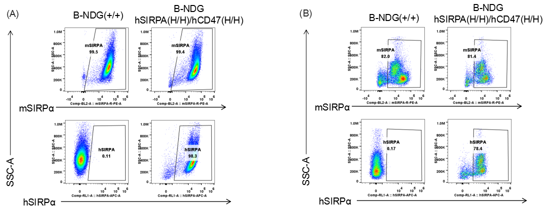
Species specific SIRPα expression analysis in B-NDG hSIRPA/hCD47 mice by flow cytometry. (A) Peritoneal lymphocyte and (B) Splenocytes from B-NDG and homozygous B-NDG hSIRPA/hCD47 mice were analyzed by flow cytometry with anti-SIRPα antibodies. Mouse SIRPα was detectable in C57BL/6, B-NDG and homozygous B-NDG hSIRPA/hCD47 mice. This anti-mouse SIRPα antibody also cross reacts with human SIRPα. Human SIRPα was exclusively detectable in homozygous B-NDG hSIRPA/hCD47 but not C57BL/6 or B-NDG mice.

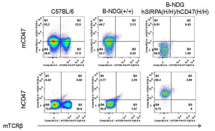
Species specific CD47 expression analysis in B-NDG hSIRPA/hCD47 mice by flow cytometry. Splenocytes from C57BL/6, B-NDG and homozygous B-NDG hSIRPA/hCD47 mice were analyzed by flow cytometry with anti-CD47 antibodies. Mouse CD47 was detectable in C57BL/6 and B-NDG mice. Human CD47 was exclusively detectable in homozygous B-NDG hSIRPA/hCD47 but not C57BL/6 or B-NDG mice.
Combination therapy of anti-human CD20 antibody with anti-human SIRPA antibody

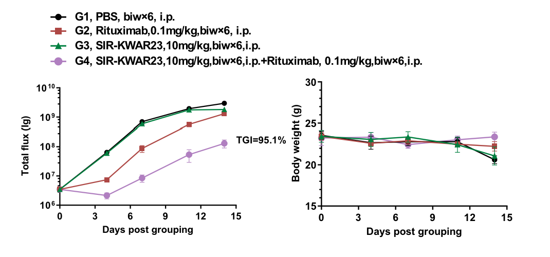
Antitumor activity of anti-human CD20 antibody with anti-human SIRPA antibody in B-NDG hSIRPA/hCD47 mice. (A) hCD20 antibody combined with hSIRPA antibody inhibited B-luciferase-GFP Raji tumor growth in B-NDG hSIRPA /hCD47 mice. Human B-luciferase-GFP Raji cells (B lymphocytes) (5.0E+05) were inoculated into homozygous B-NDG hSIRPA /hCD47 mice (female, 10-week-old, n=5). Mice were grouped when the fluorescence intensity reached approximately 3.5E6 p/sec, at which time they were treated with hCD20, hSIRPA or hCD20 plus hSIRPA antibodis (in house) with doses and schedules indicated in panel A. (B) Body weight changes during treatment. As shown in panel A, the combination of hSIRPA and hCD20 antibodies shows more inhibitory effects than individual groups. Values are expressed as mean ± SEM.
Summary
- Species specific SIRPα expression analysis in B-NDG hSIRPA/hCD47 mice by flow cytometry. Human SIRPα was exclusively detectable in homozygous B-NDG hSIRPA/hCD47 mice.
- Species specific CD47 expression analysis in B-NDG hSIRPA/hCD47 mice by flow cytometry. Human CD47 was exclusively detectable in homozygous B-NDG hSIRPA/hCD47 mice.
- Combination of hSIRPA and hCD20 antibodies shows more inhibitory effects than individual groups in B-NDG hSIRPA/hCD47 mice.


